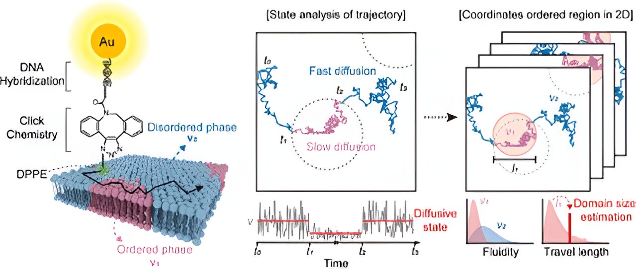

A research team led by Professor Dae-ha Seo at the Department of Physics and Chemistry, Daegu Gyeongbuk Institute of Science and Technology, has successfully developed a new technology for optical microscopy analysis—known as lipid-MAP—which can observe the microscopic phase separation in the cell membrane.
This new technology, which combines traditional microscopy with nanochemistry and machine learning, is expected to offer an important experimental strategy for exploring how cell signaling is regulated at the single-molecule level. The study is published in the journal Analytical Chemistry.
Cells, which are surrounded by the cell membrane, are the basic building blocks of all living organisms. A nanoscale, microscopic, and island-like lipid structure exists in the cell membrane. The structure plays a critical role in the interaction between biomolecules, chemical reactions, and signaling. However, it is difficult to directly observe the structure with existing observation methods.
Using gold nanoprobes and machine learning technology, a research team led by Professor Dae-ha Seo succeeded in quantitatively identifying structural properties at the nano level. Through the phenomenon of surface plasmon resonance, gold nanoparticles have the property of brightly scattering light. Using this property, the research team implemented a system that allows direct observation of the movement of single molecules by binding gold nanoparticles to lipid molecules.
By introducing an analysis technique based on machine learning algorithms, the research team detected changes in the movement of lipid molecules over a short period of time (0.01 to 0.1 seconds) and, thereby, successfully identified the micro-phase separation structure of the cell membrane.
The micro-lipid island structure, also known as the lipid raft, is known to mainly consist of cholesterol and saturated lipids, which are gathered locally. The research team confirmed that the size and properties of the structure are determined by the molecular composition of the cell membrane, and revealed that it can change based on the cholesterol content in the cell membrane or due to other various environmental factors.
Professor Seo stated, “This is the first technology to image lipid rafts in real time over a wide range, and its spatial and temporal resolution is outstanding. I hope that the research results will serve as a basis to fundamentally understand cell function and disease mechanism, and will develop into precision diagnostic technology for diseases.”
More information:
Jiseong Park et al, Analysis of Phase Heterogeneity in Lipid Membranes Using Single-Molecule Tracking in Live Cells, Analytical Chemistry (2023). DOI: 10.1021/acs.analchem.3c02655
Provided by
Daegu Gyeongbuk Institute of Science and Technology (DGIST)
Citation:
New technology for microscopy analysis provides precise maps of cell membrane lipid rafts (2023, December 11)
retrieved 11 December 2023
from https://phys.org/news/2023-12-technology-microscopy-analysis-precise-cell.html
This document is subject to copyright. Apart from any fair dealing for the purpose of private study or research, no
part may be reproduced without the written permission. The content is provided for information purposes only.


